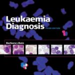
Leukaemia Diagnosis 5th Edition: The Complete Guide to Modern Leukemia Classification, Laboratory Diagnosis, and Clinical Practice
Introduction


Leukemia is one of the most extensively studied blood cancers in modern medicine. Accurate diagnosis is essential because treatment decisions, patient prognosis, and long-term survival depend heavily on identifying the exact subtype of leukemia. Advances in molecular genetics, immunophenotyping, cytogenetics, and digital pathology have transformed diagnostic approaches over the past two decades.
Leukaemia Diagnosis, 5th Edition represents one of the most respected references in hematopathology, offering comprehensive guidance for laboratory professionals, hematologists, oncologists, pathologists, medical technologists, and researchers. The book combines traditional morphology with cutting-edge molecular techniques, making it an invaluable resource for both students and experienced clinicians.
🩸 Why accurate diagnosis matters
- 🔬 Determines the exact leukemia subtype
- 💊 Guides personalized treatment
- 📊 Predicts patient prognosis
- 🧬 Identifies genetic abnormalities
- ❤️ Improves survival outcomes
- 📈 Supports precision medicine
As medicine moves toward individualized therapies, modern leukemia diagnosis increasingly relies on integrating multiple laboratory disciplines rather than depending on a single test.
Background Theory
Evolution of Leukemia Diagnosis
Historically, leukemia diagnosis depended almost entirely on microscopic examination of blood smears and bone marrow aspirates. Physicians identified abnormal white blood cells based on their size, shape, nuclear characteristics, and staining properties.
Although morphology remains important today, it no longer provides sufficient diagnostic accuracy for many leukemia subtypes.
Modern diagnosis integrates several complementary technologies:
- 🔬 Light microscopy
- 🧫 Bone marrow pathology
- 🧪 Flow cytometry
- 🧬 Cytogenetics
- 📊 Fluorescence In Situ Hybridization (FISH)
- 🧬 Polymerase Chain Reaction (PCR)
- 🧬 Next Generation Sequencing (NGS)
- 💻 Digital pathology
- 🤖 Artificial Intelligence-assisted image analysis
Each technique contributes unique information that improves diagnostic confidence.
The Importance of Integrated Diagnosis
Instead of asking:
“Does this patient have leukemia?”
Modern hematology asks:
- Which leukemia subtype?
- Which mutations are present?
- What is the patient’s risk category?
- Which targeted therapies are appropriate?
- What is the expected prognosis?
This integrated approach forms the foundation of personalized medicine.
Definition
Leukemia is a group of malignant disorders affecting the bone marrow and blood-forming tissues, characterized by uncontrolled proliferation of abnormal white blood cells.
The 5th Edition emphasizes an integrated diagnostic workflow combining:
🩸 Clinical findings
🔬 Morphology
🧪 Immunophenotyping
🧬 Cytogenetics
🧬 Molecular genetics
📈 Laboratory biomarkers
🖥️ Digital imaging
rather than relying on any single laboratory technique.
Understanding Leukemia Classification
Acute Leukemia
Acute leukemias progress rapidly because immature blast cells replace healthy bone marrow.
Examples include:
- Acute Myeloid Leukemia (AML)
- Acute Lymphoblastic Leukemia (ALL)
Symptoms often develop within weeks.
Chronic Leukemia
Chronic leukemias involve more mature blood cells and usually progress more slowly.
Examples include:
- Chronic Myeloid Leukemia (CML)
- Chronic Lymphocytic Leukemia (CLL)
Many patients remain asymptomatic for years.
Myeloid vs Lymphoid
Leukemia is also classified according to the originating cell.
| Type | Origin |
|---|---|
| Myeloid | Granulocytes, monocytes, erythroid cells |
| Lymphoid | B-cells, T-cells, NK cells |
This distinction is critical because treatment differs dramatically.
Step-by-Step Diagnostic Process
Step 1 — Clinical Assessment 👨⚕️
The diagnostic journey begins with:
- Medical history
- Family history
- Physical examination
- Symptoms assessment
Common symptoms include:
- Fatigue
- Fever
- Frequent infections
- Easy bruising
- Bleeding
- Weight loss
- Night sweats
- Bone pain
Step 2 — Complete Blood Count (CBC)
The CBC is usually the first laboratory investigation.
It evaluates:
- White blood cells
- Red blood cells
- Hemoglobin
- Platelets
Abnormal findings may include:
⬆️ Elevated white cells
⬇️ Low hemoglobin
🎯 Low platelets
Presence of blasts
Step 3 — Peripheral Blood Smear


Microscopic examination evaluates:
- Cell morphology
- Blast percentage
- Nuclear appearance
- Cytoplasmic granules
- Auer rods
- Dysplasia
Morphology remains one of the most important diagnostic tools.
Step 4 — Bone Marrow Examination
Bone marrow aspiration provides:
- Blast percentage
- Cellularity
- Fibrosis
- Dysplastic changes
Trephine biopsy offers additional structural information unavailable from aspiration alone.
Step 5 — Flow Cytometry
Flow cytometry identifies cell surface markers called CD antigens.
Examples include:
| Marker | Clinical Significance |
|---|---|
| CD34 | Stem cells |
| CD13 | Myeloid |
| CD33 | Myeloid |
| CD19 | B-cell |
| CD20 | Mature B-cell |
| CD3 | T-cell |
| CD117 | AML |
Flow cytometry often establishes lineage within hours.
Step 6 — Cytogenetic Analysis
Chromosomal abnormalities strongly influence diagnosis.
Examples include:
| Chromosome Change | Disease |
|---|---|
| t(15;17) | Acute Promyelocytic Leukemia |
| t(8;21) | AML |
| inv(16) | AML |
| Philadelphia chromosome | CML |
Step 7 — Molecular Testing
Modern laboratories routinely analyze mutations such as:
- FLT3
- NPM1
- CEBPA
- IDH1
- IDH2
- TP53
- RUNX1
- JAK2
These mutations influence:
✔ Prognosis
🎯 Drug selection
✔ Risk stratification
✔ Clinical trial eligibility
Step 8 — Integrated Final Diagnosis
The final diagnosis combines every available result into one unified report.
Comparison of Diagnostic Methods
| Method | Speed | Accuracy | Cost | Primary Purpose |
|---|---|---|---|---|
| CBC | Very Fast | Moderate | Low | Screening |
| Blood Smear | Fast | High | Low | Morphology |
| Bone Marrow | Moderate | Very High | Moderate | Confirmation |
| Flow Cytometry | Fast | Excellent | Moderate | Cell Identification |
| Cytogenetics | Slow | Excellent | High | Chromosomes |
| PCR | Fast | Excellent | Moderate | Mutation Detection |
| NGS | Moderate | Outstanding | High | Comprehensive Genetics |
Diagnostic Workflow Diagram
Patient Symptoms
       │
       ▼
Physical Examination
       │
       ▼
CBC
       │
       ▼
Blood Smear
       │
       ▼
Bone Marrow
       │
       ▼
Flow Cytometry
       │
       ▼
Cytogenetics
       │
       ▼
Molecular Testing
       │
       ▼
Integrated Diagnosis
       │
       ▼
Treatment Plan
Examples
Example 1
A 35-year-old patient presents with fatigue and bruising.
CBC reveals:
- WBC = 55,000
- Hemoglobin = 8.4
- Platelets = 40,000
Bone marrow shows 80% blasts.
Flow cytometry demonstrates:
- CD13+
- CD33+
- CD34+
Diagnosis:
✅ Acute Myeloid Leukemia
Example 2
A 10-year-old child presents with fever and recurrent infections.
Peripheral blood reveals lymphoblasts.
Flow cytometry:
- CD19+
- CD10+
- TdT+
Diagnosis:
✅ Acute Lymphoblastic Leukemia
Example 3
A routine blood test reveals WBC = 120,000.
PCR confirms BCR-ABL1.
Diagnosis:
✅ Chronic Myeloid Leukemia
Real-World Applications
🏥 Hospital hematology laboratories
🧪 Clinical pathology laboratories
🧬 Molecular diagnostic centers
🎓 Medical schools
🔬 Cancer research institutes
💻 Artificial Intelligence research
📈 Pharmaceutical clinical trials
🌍 National cancer screening programs
Common Mistakes
❌ Depending only on CBC
❌ Ignoring morphology
🎯 Poor bone marrow sampling
❌ Delayed molecular testing
❌ Misinterpreting flow cytometry
🎯 Incorrect specimen handling
❌ Failure to integrate laboratory findings
❌ Delayed referral to specialists
Challenges and Solutions
| Challenge | Solution |
|---|---|
| Similar leukemia subtypes | Multi-modal testing |
| Limited resources | Prioritize essential investigations |
| Delayed diagnosis | Standardized diagnostic pathways |
| Genetic complexity | Next Generation Sequencing |
| Rare mutations | Reference laboratories |
| Human interpretation errors | Digital pathology and AI |
Case Study
Patient Profile
👨 Male
Age: 47 years
Symptoms:
- Persistent fatigue
- Fever
- Easy bruising
- Gum bleeding
Laboratory Results
CBC
- WBC: 68,000
- Hemoglobin: 7.9 g/dL
- Platelets: 32,000
Peripheral smear:
- Numerous blasts
- Auer rods
Bone marrow:
- 72% myeloblasts
Flow cytometry:
- CD34
- CD117
- CD13
- CD33
Cytogenetics:
- t(8;21)
Molecular testing:
- RUNX1-RUNX1T1 fusion
Final Diagnosis
Acute Myeloid Leukemia with favorable-risk cytogenetics.
Outcome
Following induction chemotherapy and risk-adapted consolidation therapy, the patient achieved complete remission. Continuous molecular monitoring enabled early detection of minimal residual disease and informed follow-up decisions.
Tips for Doctors
🩺 Never rely on a single laboratory result.
🔬 Correlate morphology with immunophenotyping.
🧬 Request molecular testing early when acute leukemia is suspected.
📊 Use standardized classification systems such as the latest WHO and ICC recommendations.
🤝 Encourage multidisciplinary collaboration among hematologists, pathologists, molecular geneticists, and laboratory scientists.
📁 Maintain comprehensive documentation to support treatment planning and future comparisons.
🎯 Monitor measurable (minimal) residual disease when appropriate to assess treatment response and detect relapse early.
Frequently Asked Questions (FAQs)
1. Why is bone marrow examination necessary?
Bone marrow examination confirms the diagnosis, determines the percentage of blast cells, evaluates marrow architecture, and provides material for cytogenetic and molecular analyses.
2. Can leukemia be diagnosed from a blood test alone?
Sometimes peripheral blood findings strongly suggest leukemia, but a complete diagnosis usually requires bone marrow studies along with immunophenotyping and genetic testing.
3. What is flow cytometry used for?
Flow cytometry identifies the immunophenotype of abnormal cells by detecting specific cell-surface and intracellular markers, helping determine the leukemia lineage and subtype.
4. Why are genetic tests important?
Genetic abnormalities influence prognosis, help classify the disease more precisely, guide targeted therapies, and support risk stratification.
5. What is the Philadelphia chromosome?
It is a chromosomal translocation, t(9;22), that creates the BCR-ABL1 fusion gene. It is classically associated with Chronic Myeloid Leukemia and is also found in some cases of Acute Lymphoblastic Leukemia.
6. Is artificial intelligence replacing hematopathologists?
No. AI serves as a decision-support tool by assisting with image analysis, pattern recognition, and workflow efficiency, while final interpretation remains the responsibility of qualified medical professionals.
7. How has the 5th Edition improved leukemia diagnosis?
It emphasizes integrated diagnostics, incorporating morphology, immunophenotyping, cytogenetics, molecular genetics, and contemporary disease classification to support precision medicine.
Conclusion
The Leukaemia Diagnosis, 5th Edition reflects the modern evolution of hematologic diagnostics, where accurate classification depends on combining clinical assessment with advanced laboratory technologies. While traditional microscopy remains indispensable, today’s diagnostic process also relies on flow cytometry, cytogenetics, molecular testing, and increasingly, digital pathology and genomic analysis.
For students, the text provides a strong foundation in the principles of leukemia diagnosis. For laboratory professionals and clinicians, it serves as a practical reference for applying standardized diagnostic workflows and interpreting complex findings. As precision medicine continues to advance, integrated diagnostic strategies remain central to selecting appropriate therapies, improving prognostic accuracy, and ultimately achieving better outcomes for patients with leukemia.